Digital Dental X-Rays in Irmo

Modern Dental Imaging Technology
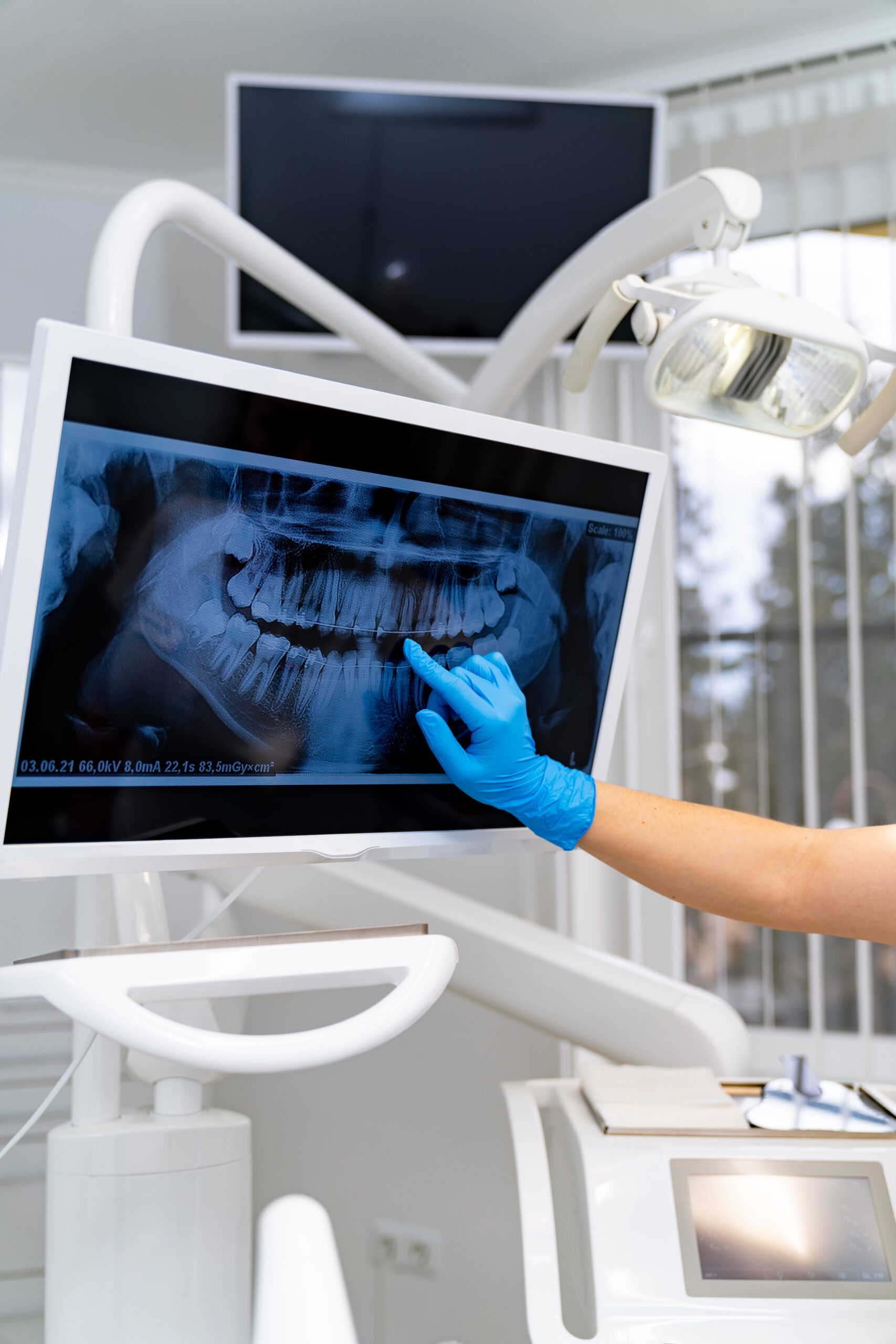
Digital X-rays represent a significant advancement in dental diagnostics, providing detailed images of your teeth, roots, and surrounding bone structure with greater precision than traditional film X-rays. This state-of-the-art imaging technology is a pillar of exceptional preventive dental care as it allows our team to detect problems in their earliest stages, when treatment is most effective and least invasive.
Our digital imaging system produces high-quality images instantly, eliminating the wait time associated with film processing. The enhanced image quality allows for better diagnosis and treatment planning, while the digital format enables us to easily share images with specialists when needed and store them securely in your electronic health record.
Dental emergencies happen. Get immediate professional care with our same-day emergency appointment availability.

Benefits of Digital X-rays
Reduced Radiation Exposure
Digital X-rays require up to 80% less radiation exposure compared to traditional film X-rays, making them significantly safer for patients of all ages. The advanced sensor technology captures detailed images with minimal radiation, providing excellent diagnostic quality while prioritizing your health and safety during routine dental examinations and specialized imaging procedures.
Instant Results and Diagnosis
Digital images appear on our computer screen immediately after exposure, eliminating the time-consuming film development process. This instant availability allows us to review your X-rays with you during your appointment, explain findings clearly, and begin treatment planning right away. The immediate results improve efficiency and enhance your understanding of your oral health status.
Enhanced Image Quality
Digital sensors capture incredibly detailed images that can be magnified and enhanced for better diagnosis. We can adjust contrast, brightness, and magnification to examine specific areas more closely, leading to more accurate diagnoses and comprehensive treatment planning. The superior image quality helps detect problems that might be missed with traditional X-ray methods.
Digital X-ray Process
Process
Step 1
Preparation and Positioning
The digital X-ray process begins with comfortable positioning using specialized sensors placed in your mouth. These sensors are smaller and more comfortable than traditional film, reducing discomfort during the imaging process. Our team ensures proper positioning for optimal image quality while maintaining your comfort throughout the procedure.
Step 2
Image Capture
The digital sensor captures detailed images of your teeth and supporting structures in seconds. The advanced technology requires minimal radiation exposure while producing high-resolution images that reveal even the smallest details of your oral health. Multiple angles may be captured to provide a comprehensive view of your dental condition.
Step 3
Immediate Review and Analysis
Your digital X-rays appear instantly on our computer monitor, allowing immediate review and analysis. We can examine the images in detail, looking for signs of decay, infection, bone loss, or other dental problems. The digital format allows for easy comparison with previous X-rays to monitor changes over time.
Step 4
Treatment Planning and Discussion
Using the detailed digital images, we develop comprehensive treatment recommendations tailored to your specific needs. The clear images help us explain any problems we discover and discuss treatment options with you. Digital X-rays enable more accurate treatment planning and better patient education about oral health conditions.
Step 5
Image Storage and Sharing
Your digital X-rays are stored securely in our electronic records system, making them easily accessible for future appointments and treatment planning. When referrals to specialists are needed, digital images can be shared instantly, improving coordination of care and eliminating the need for retaking X-rays at different offices.

Comprehensive Diagnostic Capabilities
Digital X-ray technology enables the detection of a wide range of dental conditions that may not be visible during a routine visual examination. These advanced imaging capabilities allow our team to identify problems in their earliest stages, when treatment is typically more conservative and successful.
The detailed images help us evaluate tooth structure, root health, bone density, and the position of developing teeth. This comprehensive diagnostic capability is essential for maintaining optimal oral health and preventing more serious problems from developing over time.
Dr. Jameson Davis - Digital Imaging
Dr. Davis utilizes the latest digital X-ray technology to provide the most accurate diagnoses and comprehensive treatment planning for his patients. His expertise in digital imaging interpretation ensures that even the smallest dental problems are detected early, when treatment is most effective. Dr. Davis takes time to review digital X-rays with each patient, explaining any findings clearly and discussing how they relate to overall oral health. His commitment to using advanced diagnostic technology reflects his dedication to providing the highest quality dental care available.

FAQS
How often do I need digital X-rays?
The frequency of X-rays depends on your individual oral health needs and risk factors. Most patients require routine X-rays once a year, while those with active dental problems or higher risk factors may need them more frequently. We follow established guidelines and only recommend X-rays when clinically necessary for diagnosis and treatment planning.
Are digital X-rays safe during pregnancy?
While digital X-rays use significantly less radiation than traditional film, we typically postpone routine X-rays during pregnancy unless there’s a dental emergency requiring immediate diagnosis. If X-rays are necessary, we use additional protective measures, including lead aprons, to ensure the safety of both mother and baby.
How do digital X-rays compare to traditional film X-rays?
Digital x-rays offer superior image quality with up to 80% less radiation exposure compared to traditional film. They produce instant results, are more environmentally friendly, and can be easily stored and shared. The enhanced diagnostic capabilities and improved patient experience make digital X-rays the preferred choice for modern dental care.
Can digital X-rays detect all dental problems?
While digital X-rays are excellent diagnostic tools, they cannot detect all dental conditions. Some problems, such as early gum disease or surface cavities, are better detected through visual examination and other diagnostic methods. Digital X-rays are most effective when combined with comprehensive clinical examination and other diagnostic tools.
What happens if a problem is found on my X-ray?
If we discover any issues on your digital X-rays, we’ll discuss the findings with you immediately and explain the recommended treatment options. The detailed images help us provide accurate diagnoses and develop effective treatment plans. We’ll review the images with you so you understand the condition and the importance of recommended treatment.
Do you keep copies of my digital X-rays?
Yes, all digital X-rays are stored securely in your electronic health record and are available for future reference and treatment planning. This digital storage system ensures your images are safely preserved and easily accessible when needed. We can also provide copies for your records or when transferring to another dental office.
